News (Good News Only!)

    - March, 2024: Our new paper on proposing a new boundary loss function for image segmentation is now published by Information Sciences. "Boundary-wise loss for medical image segmentation based on fuzzy rough sets", Information Sciences.

    - March, 2024: Three papers were accepted by IEEE-ISBI 2024,. Congratulations to the students. They will present the works in Athens in May.
    • Benjamin Towle, Xin Chen and Ke Zhou, "SimSAM: Zero-shot Medical Image Segmentation via Simulated Interaction". accepted by IEEE-ISBI, oral presentation, 2024.
    • Zhendi Gong, Andrew French, Guoping Qiu and Xin Chen, "CTrans: A Multi-resolution Convolution-transformer Network for Medical Image Segmentation." accepted by IEEE-ISBI, oral presentation, 2024.
    • Ruizhe Li, Dorothee Auer, Christian Wagner and Xin Chen, " MrRegNet: Multi-resolution Mask Guided Convolutional Neural Network for Medical Image Registration with Large Deformations", accepted by IEEE-ISBI, 2024.


Old News

- October, 2023: Huge congratulations to Qiao Lin, who successfully defended her PhD thesis: "Incorporating Fuzzy-based Methods to Deep Learning Models for Semantic Segmentation" subject to minor corrections! A few more upcoming papers from her thesis.

- September, 2023: Many congratulations to Ruizhe Li who passed his PhD viva subject to minor corrections! The title of his thesis is "Semi-supervised Learning for Medical Image Segmentation". He has also been awarded the University Tri Campus Postgraduate Prize: Andrew Hendry Postgraduate Scholarship, as a recognition of the contributions he has made in his research.

- August, 2023: Our new paper on out-of-distirbution detection has been published in Fuzz-IEEE conference. "Fuzzy Uncertainty-based Out-of-distribution Detection Algorithm for Semantic Segmentation", IEEE International Conference on Fuzzy Systems 2023.

- March, 2023: I am now Associate Professor of Computer Science

- January, 2023: Now our grand challenge winner paper has been published "Motion-Related Artefact Classification Using Patch-Based Ensemble and Transfer Learning in Cardiac MRI" International Workshop on Statistical Atlases and Computational Models of the Heart, MICCAI 2022. ArXiv version

- December, 2022: Congrats to Qiao's paper "A Novel Quality Control Algorithm for Medical Image Segmentation Based on Fuzzy Uncertainty" has now been published by IEEE Transactions on Fuzzy Systems. Well done!

- October, 2022: as one of the co-Is, our multi-million project that aims to measure, model and predict gestational development was funded by Wellcome Leap's In Utero programme.

- September, 2022: There is nothing happier than witnessing a talented and diligent PhD student (Ruizhe Li) get rewarded! We are the winner of task 1 CMRxMotion Challenge held in MICCAI2022 .


- July, 2022: Mina Jafari's paper "LMISA: A Lightweight Multi-modality Image Segmentation Network via Domain Adaptation using Gradient Magnitude and Shape Constraint" has now been published by Medical Image Analysis. Well done!

- June, 2022: Many congratulations to Mina Jafari on successfully defended her PhD thesis subjective to minor corrections, entitled "Improvement of Model Performance and Generalisability of Deep Convolutional Neural Networks for Medical Image Segmentation." She is now working as a Post-doc researcher at Imperial College London. All the best!

- June, 2022: Huge congrats to Zixiao, who is now officially Dr. Shen. He has been awarded his PhD on the topic of feature selection using Fuzzy-based methods.

- June, 2022: Congrats to Zhendi's first paper, which is accepted by MIUA 2022. The paper presents a muscle segmentation method on Whole-body MRI based on his MSc work. He is now a year 1 PhD student under my supervision.

- May, 2022: Qiao Lin's new paper on "Quality Quantification in Deep Convolutional Neural Networks for Skin Lesion Segmentation using Fuzzy Uncertainty Measurement" has been accepted by IEEE-WCCI 2022 as an oral presentation.

- April, 2022: Golnar K. Mahani's new idea for bounding box based image segmentation is published in IEEE-ISBI 2022. Bounding Box Based Weakly Supervised Deep Convolutional Neural Network for Medical Image Segmentation Using an Uncertainty Guided and Spatially Constrained Loss.

About

I am an Associate Professor of Computer Science at the University of Nottingham, UK. My research interests are image processing, computer vision and machine learning, particularly applied to medical image analysis. My team and I develop algorithms for 2D/3D image segmentation, image registration, statistical shape/ motion modelling, classification/ regression models and CT & MRI image reconstruction, which have been successfully applied to different medical applications (e.g. breast cancer, diabetes care, wrist injury and radiotherapy). I am an active member in the computer vision and medical imaging community, regularly serve as reviewers for international conferences and prestigious journals (e.g. Medical Image Analysis, MICCAI, IEEE-ISBI, BMVC, IEEE-TMI, IEEE-TBME, Physics in Medicine and Biology, etc.).

Previous Projects

The wrist is one of the most complex and vulnerable joints in the body, consisting of eight carpal bones. Wrist pain is currently diagnosed by expert assessment of abnormal carpal bone movements in 2D fluoroscopy sequences. The overall aim of the current project is computer interpretation of these 2D sequences to recover the 3D motion of the carpal bones, and further leads to quantitative measurements of wrist diseases.

- Chen, X., et al., Automatic Inference and Measurement of 3D Carpal Bone Kinematics From Single View Fluoroscopic Sequences. IEEE Transactions on Medical Imaging, 2013. 32(2): p. 317-328.

- Chen, X., et al., Automatic Generation of Statistical Pose and Shape Models for Articulated Joints. IEEE Transactions on Medical Imaging, 2014. 33(2): p. 372 - 383.

- Chen, X., et al., Inferring 3D kinematics of carpal bones from single view fluoroscopic sequences, MICCAI 2011, Springer. p. 680-687.

- Chen, X., et al., Integrated frameworkfor simultaneous segmentation and registration of carpal bones, ICIP 2011, p. 433-436.

Corneal confocal microscopy is a novel in-vivo imaging modality that has the potential to be a non-invasive and objective image biomarker for peripheral neuropathy. Automatic quantification of nerve morphology is a major step forward in the early diagnosis and assessment of progression, and, in particular, for use in clinical trials to establish therapeutic benefit in diabetic and other peripheral neuropathies.

- Chen, X., et al., An Automatic Tool for Quantification of Nerve Fibers in Corneal Confocal Microscopy Images. IEEE Transactions on Biomedical Engineering, 2017. 64(4): p. 786-794.

- Chen, X., et al., Small Nerve Fiber Quantification in the Diagnosis of Diabetic Sensorimotor Polyneuropathy: Comparing Corneal Confocal Microscopy With Intraepidermal Nerve Fiber Density. Diabetes Care, 2015. 38(6): p. 1138-1144.

Currently, breast screening is almost exclusively performed with mammography. However, for women with dense breasts the sensitivity of mammography for detecting breast cancer is low. The aim of the project is to develop methods to personalise breast cancer screening, based on risk and breast density markers. We have deleloped method to estimated volumetric breast density from single view mammography.

- Chen, X., et al., Improving Mammographic Density Estimation in the Breast Periphery, IWDM 2016, p. 469-477

- Chen, X., et al., A Novel Framework for Fat, Glandular Tissue, Pectoral Muscle and Nipple Segmentation in Full Field Digital Mammograms, IWDM 2014. p. 201-208

- Chen, X., et al., Breast Cancer Risk Analysis Based on a Novel Segmentation Framework for Digital Mammograms, MICCAI 2014. p. 536-543.

Dynamic magnetic resonance imaging (MRI) involves imaging a region of interest with high temporal resolution, and is useful in many applications in which knowledge of motion is of interest. We present a novel retrospective self-gating method based on manifold alignment (MA), which enables reconstruction of free breathing, high spatial, and temporal resolution abdominal magnetic resonance imaging sequences.

- Chen, X., et al., High-Resolution Self-Gated Dynamic Abdominal MRI Using Manifold Alignment. IEEE Transactions on Medical Imaging, 2017. 36(4): p. 960 - 971.

- Chen, X., et al., Efficient deformable motion correction for 3-D abdominal MRI using manifold regression, MICCAI 2017, p. 270-278.

- Chen, X., et al., Dynamic Volume Reconstruction from Multi-slice Abdominal MRI Using Manifold Alignment, MICCAI 2016, p. 493-501.

Teaching

Software Tools

Image segmentation is a crucial step in many medical image analysis processes. Manual or semi-automatic image segmentation is often necessary to provide accurate annotations for supervised machine learning algorithms or to be directly used for clinical feature quantification. Our software aims to enable rapid interactive image segmentation for both 2D and 3D medical images based on full-connected conditional random field method. It supports up to 10 foreground labels and various image format (Matlab, Nifty, DICOM, etc.). The software was developed in Matlab, hence the Matlab runtime library will be automatically installed. Please make sure you have internet connections during the installation process. The software is freely available for research purposes, please cite our paper if it is useful to your project.

Paper

Software download

Video instruction of the software.

Corneal confocal microscopy is a novel in-vivo imaging modality that has the potential to be a non-invasive and objective image biomarker for peripheral neuropathy. Automatic quantification of nerve morphology is a major step forward in the early diagnosis and assessment of progression, and, in particular, for use in clinical trials to establish therapeutic benefit in diabetic and other peripheral neuropathies.
The software has been recently updated using deep learning method. Simply download the tool from the link below, unzip it to your local drive, click "CCMAnalysis.exe" to run (no installation needed). It currently only support Windows machines.
If you find it useful, please cite our paper: N. Zhang et al., A spatially constrained deep convolutional neural network for nerve fiber segmentation in corneal confocal microscopic images using inaccurate annotations. IEEE-ISBI, 2020

Software download

Research Grants

- 2022-2026 Wellcome Leap funded, Co-investigator, “Maternal venous return from the placenta and the effect of placental and uterine contractions as potential markers of stillbirth risk”.

- 2021-2025 MRC funded, Co-investigator, “DEMISTIFI Multi Morbidity: DEfining MechanIsms Shared across mulTI-organ FIbrotic disease to prevent the development of long term multi-morbidity”.

- 2021-2025 ERC funded, Co-investigator, “Brain connectivity metrology for personalised neuroimaging in health and disease”.

- 2021-2022 NIHR/NHSx AI Award, Co-lead, “Personalised Preoperative Neoadjuvant Chemotherapy to Optimise Curative Treatment in Breast Cancer”.

- 2020-2022 Weston Brain Institute Award, Co-investigator, “Determining subtype-specific rates of brainstem depigmentation as progression marker in early Parkinson’s: A serial neuromelanin MRI study to inform stratified trial designs”.

- 2020-2022 British Heart Foundation Award, Co-investigator, “Assessment of artificial neural networks and conventional statistical regression techniques in diagnosis and prediction of outcome after stroke using big data”.

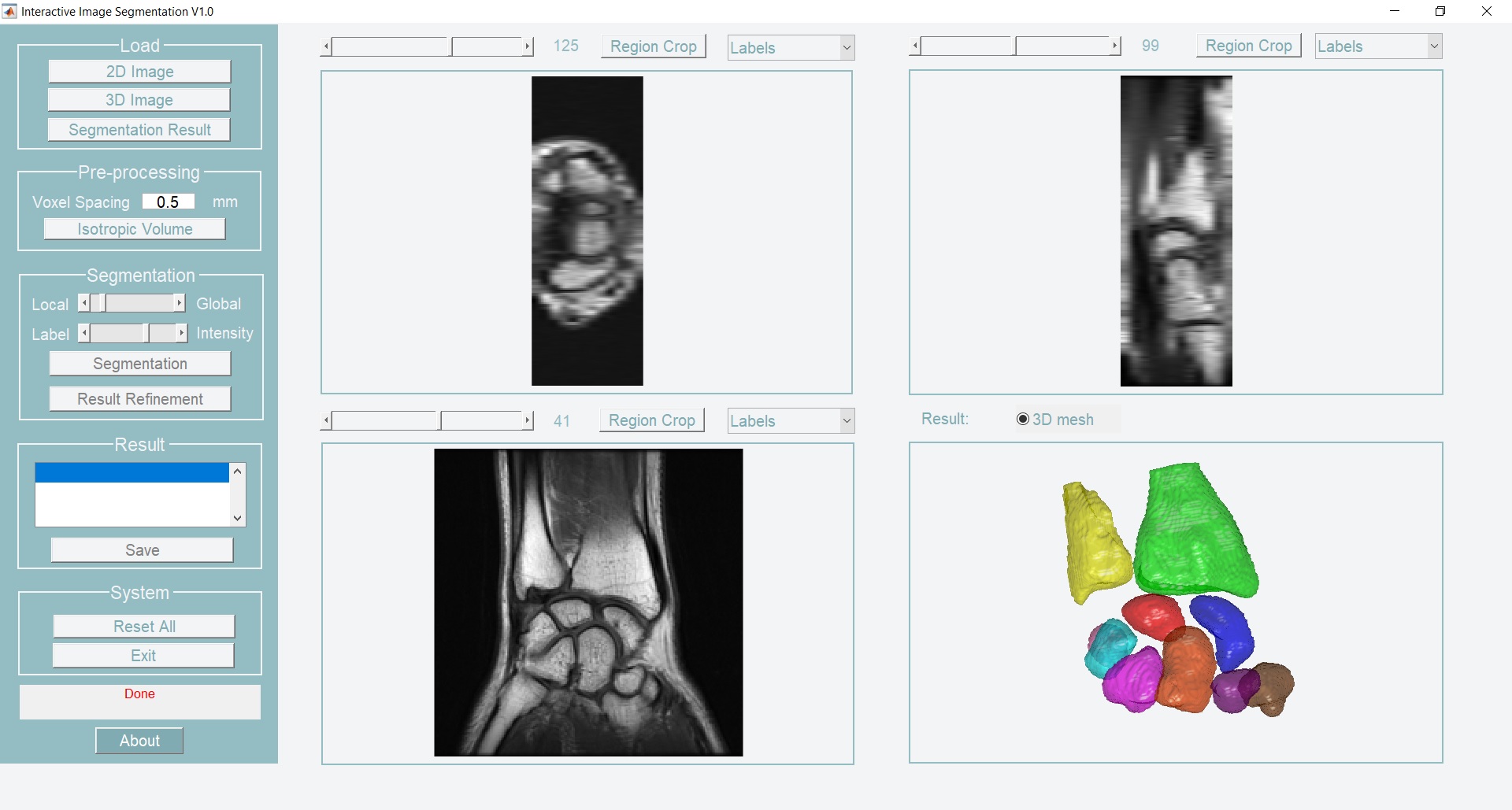
- 2019-2021 Precision Imaging Beacon Catalyst Award by Precision Imaging Beacon, University of Nottingham, Co-investigator, “Developing an artificial intelligence augmented diagnostic imaging strategy for patients with occult scaphoid fractures of the wrist”.

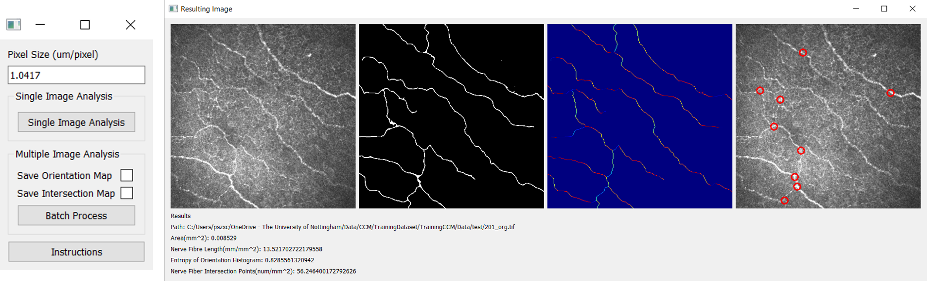
- 2018-2019 Research Accelerator Award by Precision Imaging Beacon, University of Nottingham, Principle investigator, “Automated blood vessel quantification for 7 Tesla magnetic resonance angiography”.

Team Members

Mina Jafari

PhD graduated in 2022

RuiZhe Li

PhD graduated in 2023

Qiao Lin

PhD graduated in 2023

Golnar Mahani

PhD graduated in 2024

Zhendi Gong

PhD student (12/2021- )

Benjamin Towle

PhD student (10/2021-)

Stephen Lloyd-Brown

PhD student (10/2022- )

Boya Wang

PhD student (12/2022- )

Rongjun Dong

PhD student (10/2023-)

Xinyi Wang

PhD student (10/2023-)